


Confocal Microscopy Laboratory
Calendars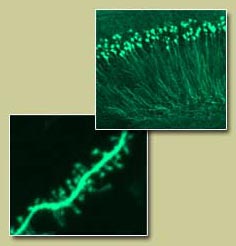
Zeiss LSM 880
The Zeiss LSM 880 is an inverted microscope. Configuration includes 405,458,488,514,543,594,and 633nm lasers. It is has additional Modules for Tiles & Positions, FRAP, 3D VisArt, FCCS and RICS. Also included temperature and CO2 control chamber for live cell imaging.
Objectives available:
EC Plan-Neofluar 10x/0.30 WD=5.2 M27
Plan-Apochromat 20x/0.8 WD=0.55 M27
Plan-Apochromat 63x/1.40 Oil DIC M27
alpha Plan-FLUAR 100x/1,49 Oil (UV-VIS)
The Zeiss LSM 880 is located in room 2311.
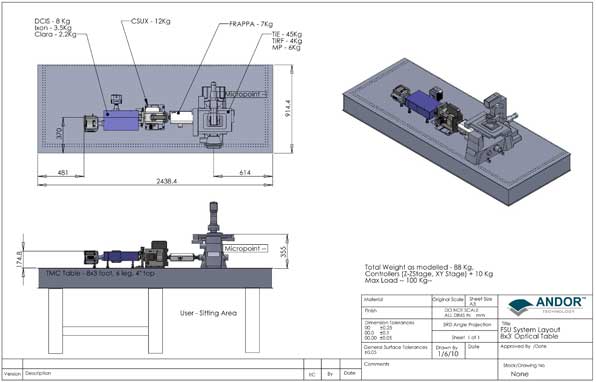
Andor Revolution Spinning Disk Laser Confocal Microscope for Live Cell Imaging
Consists of the following:
- Yokogawa automated 5000rpm spinning disk
- Nikon Eclipse Ti Microscope with perfect focus system for automatic focus drift correction to enable long term imaging
- Andor Revolution system with FRAPPA(Fluorescence Recovery After Photobleaching and Photo Activation)
- Total 7 lasers solid state laser lines (405nm, 44nm5, 488nm, 515nm, 561nm, 640nm) plus a dye based pulsed nitrogen laser capable of generating multiple wavelengths from true UV (364nm) to [Micropoint system from Photonics Instruments] for UV uncaging, photoablation and laser induced DNA damage (DNA damage will also be carried out using the 405nm solid state laser and Micropoint 539nm dye based pulsed nitrogen laser). The 405nm and the 488nm lasers are powerful at 100mW for photobleaching (FRAP/FLIP), photoconversion, photoactivation and FRET.
- Total Internal Reflection Fluorescence (TIRF) microscopy
- High NA objectives of the following magnifications: 20X, 40X oil, 60X water, 60XTIRF oil, 100XTIRF oil and a 100X oil with high UV transmission. DIC for all the objectives.
- Dual camera set up (one Andor 897 EMCCD for high speed and high sensitivity, the other Andor Clara for high resolution)
- Piezo-Z for fast Z-stacks
- Stage to accommodate glass bottom 35mm dishes and multi well plates
- Stage top incubator and CO2 control/ gas mixer unit that allows CO2 concentrations of up to 10% and hypoxia conditions down to 0.1% O2. Plus any necessary adaptors to accommodate the dishes, multiwell plates and slides.
- Optimal filter/dichroic sets for blue, cyan, green, yellow, red and far red fluorescence
- 6 wavelength LUMENCOR based illumination system for the microscope (instead of traditional mercury or metal halide lamps for use when lasers are not required for imaging).
- Andor iQ image acquisition software
- Bitplane Imaris 64-bit image analysis/deconvolution software with site license
- Two high end 64-bit computer workstations for handling large files (one workstation for image acquisition, the other for analysis) with high resolution monitors
- Anti-vibration table
OMX Blaze
The DeltaVision OMX offers super-resolution imaging using 3D structured illumination (3D-SIM) and/or localization microscopy techniques. The Blaze SIM Module generates an illumination pattern in three dimensions giving a twofold resolution improvement in the X, Y and Z. This translates into an eight fold increase for a 3D volume. The Blaze design consists of an opto-mechanical light pattern generator incorporating high speed galvanometers for manipulating the light beams necessary to generate the 3D-SIM pattern. This is essential as simple line gratings cannot generate the necessary pattern to allow full three dimensional super resolution reconstruction or fully optimize for each excitation wavelength.
Three-Dimensional Structured Illumination (3D-SIM)
3D-SIM is a super-resolution technique that uses structured illumination patterns to achieve sub-diffraction limit imaging. The structured light pattern creates interference moiré patterns with the fine structures of the sample which are captured in the resulting fluorescence images. The sample is imaged using several different orientations and phases of the light pattern then computer algorithms process the data set and generate the final super-resolution image.
Emission filters:
- DAPI or equivalent dye 435/31 nm
- CFP or equivalent dye 477/35 nm
- GFP,AlexaFluor®488 or equivalent 528/48
- YFP or equivalent dye 541/22 nm
- mCherry, AlexaFluor® 568 or equivalent dye 609/37 nm
- Cy5/AlexaFluor® 647 or equivalent dye 683/40 nm
Resolution:
- 100-160 nm lateral (depending on wavelength and optics)
- 300-400 nm axial (depending on wavelengths and optics)
- It is capable of imaging events in live cells at super resolution.
- Capable of 3D imaging at 1 micron slice (120 images) in <1 sec (depending on exposure time)
- Capable of 2D-SIM imaging at 20 reconstructed frames per second
- Exclusive high speed galvanometer controller laser shutters with ~200 ?sec response time
Lasers:
405 nm, 488 nm, 568 nm and 642 nm
Multi- Camera Acquisition:
Can acquisition up to 4 cameras without switching any optical elements, filters or prisms. Multiple cameras operating simultaneously enable imaging of multiple fluorescent probes with no time delays which is critical for applications such as high speed widefield live cell imaging or FRET imaging.
- Configured with four custom liquid cooled sCMOS cameras
- Simultaneous multichannel or sequential acquisition modes user selectable through software
- Custom designed optical path with high scatter light suppression systems for low background imaging noise
- Each camera channel correctable for axial chromatic shift, focus, rotation and magnification to ensure all detectors are optimally aligned for sample
High Speed Widefield Imaging:
In addition to its super-resolution capability, the DeltaVision OMX system is capable of high speed widefield imaging a solid state illumination system.
- Imaging speeds over 400 frames per second, simultaneously on 4 channels
- Exclusive 6 color solid state illumination system capable of running 4 simultaneous excitation wavelengths. Nominal excitation wavelengths 405nm, 445nm, 488nm, 514nm, 568nm, 642nm
Entire System Environmental Enclosure:
The entire DeltaVision OMX microscope is enclosed in a special cabinet that helps control temperature fluctuations, room airflow vibrations and acoustic noise that are critical when imaging at the sub-micron level. This also allows the system to be operated in any room environment eliminating the need to darkrooms or special facilities.
- Class 1 Laser safe device with laser safety interlock shutters and warning indicators
- Entire system enclosure enables operation in a normal laboratory environment, no darkroom needed
- HEPA air filtration with internal positive pressure environment to minimize dust contamination of optical surfaces
Hardware Autofocus:
The laser based hardware autofocus enables our exclusive focus finding feature that can quickly bring a sample to focus then lock on to track the focal position long term.
- Laser based hardware autofocus
- Exclusive FocusAssist system for rapid acquisition of focal plane
- Seamless integration into specimen location and scanning workflow with Spiral Mosaic automated “pan and scan” function
Charges
| Equipment | Internal | External |
|---|---|---|
Zeiss_LSM880- Equipment Use and Analysis per hour | $100 | $170 |
| Live Cell Imaging Confocal - Equipment Use and Analysis per hour | $70 | $119 |
| Live Cell Imaging Confocal - Equipment Use and Analysis per 1/4 day (4-11 hours) | $80 | $136 |
| Live Cell Imaging Confocal - Equipment Use and Analysis per 1/2 day (12-19 hours) | $100 | $170 |
| Live Cell Imaging Confocal - Equipment Use and Analysis per day (19+ hours) | $140 | $238 |
| Live Cell Imaging Confocal - Equipment Use and Analysis per week (multi day use- up to 170 hours) | $307.56 | $522.85 |
| OMX_Super Resolution - Equipment Use and Analysis per hour | $80 | $136 |
Frequent users have access to microscope one continuous week per month as well as intermittent periods up to one continuous week as scheduling permits. A commitment of 1 year is asked of frequent users.
**Scheduling available up to 1 month in advance.