By: Patience Moseley
FSU College of Medicine
Roughly 20 million people receive a cancer diagnosis each year, and an estimated 1-in-5 people will receive a cancer diagnosis in their lifetime. Cancer cells arise from damage to the cell’s instruction manual – DNA.
Our cells contain natural repair machinery that, under normal conditions, can cut out the damaged DNA and fill in the blanks to provide working instructions, preventing healthy cells from becoming cancer cells. Failure to repair DNA damage can lead to cancer cell growth and division. In fact, many cancer cells use the same, but deficient, repair machinery. When these repair systems do not function properly, DNA damage can persist and additional mutations can accumulate.
Researchers in the Department of Biomedical Sciences at Florida State University’s College of Medicine are changing the way scientists think about the DNA repair process, paradoxically, a key player in both cancer prevention and growth.
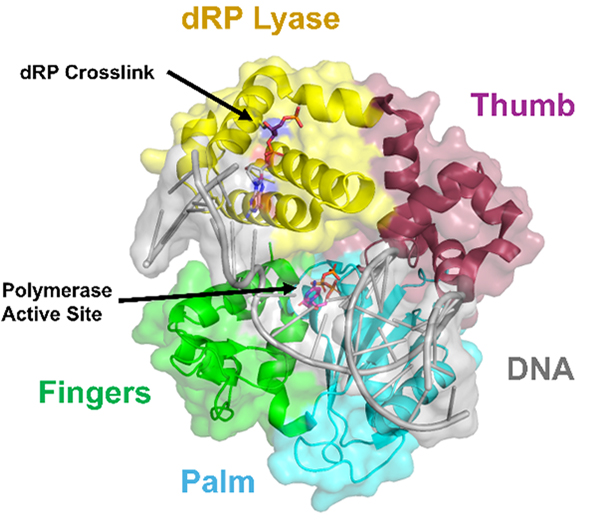
For decades, scientists thought that polymerase beta (Polβ), one of the main players in the repair machinery, would form multiple temporary bonds with the DNA during the process.
It was thought that Polβ would bind to the DNA strand, cut out the incorrect sequence, break the bond, and bind again to fill in the blanks.
It seems that may not be the case.
One research team, led by Eminent Professor and Dorian & John Blackmon Chair in Biomedical Science, Zucai Suo, Ph.D. has discovered that Polβ forms a chemical bond with DNA, and proceeds with cutting and repairing the damaged DNA while covalently bonded, or crosslinked, to the strand. By remaining attached during this critical stage of repair, the enzyme is less likely to disengage before the repair process is complete.
“We found that crosslinked Polβ actually enhances the flux — so, basically, enhances its likelihood to move forward and complete the reaction,” said Daniel Betancourt, a doctoral candidate and the lead author on the team’s publication in Nucleic Acids Research. “So that changes a lot; Polβ has only really been studied when the DNA is not covalently linked to the enzyme.”
Using these findings together with X-ray crystallography snapshots, the Suo lab established a kinetic model that outlines the different step-by-step process for DNA repair while chemically attached to the strand. The model revealed that important repair activities occur while the enzyme remains covalently bound to DNA, a state that had received relatively little attention in previous studies. In this case, Polβ doesn’t detach, or dissociate, after the initial bond is formed; it’s work is done while it’s bound to the DNA.
This work, published in June 2026, provides a wider-angle view of DNA damage and repair and lays the foundation for future research to explore how this process occurs naturally and how different forms of DNA damage might influence the repair process.
With these findings in mind, the Suo lab hopes to determine how the initial crosslinking reaction between Polβ and DNA occurs and how that bond sets the stage for successful DNA repair. Their work also establishes a foundation for future studies of at least five other human DNA polymerases involved in DNA repair and damage bypass.